Clinical Case 8
When Perfect Symmetry Is Not the Right Treatment
Fixed rehabilitation in a patient with a repaired cleft palate, prioritizing proportion, function, and cleansability over an idealized midline
In this case, the patient was a 20-year-old woman with a history of repaired cleft palate who had lost several teeth adjacent to the cleft region. For a young patient, the problem was not simply a few missing teeth; esthetics, function, and a natural-looking smile all mattered at the same time. The remaining anatomy also meant that the case could not be approached using the same assumptions applied to a conventional dental arch.
Because of the conditions in the cleft region, implant placement in that area was not possible. A tooth-supported fixed solution was therefore selected: a five-unit bridge supported by teeth 12 and 23, with three pontics between them.
The main problem
It was not simply about replacing three teeth
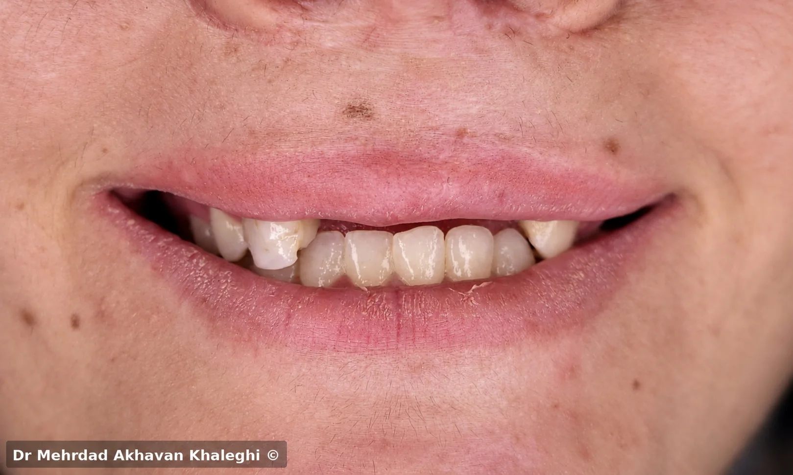
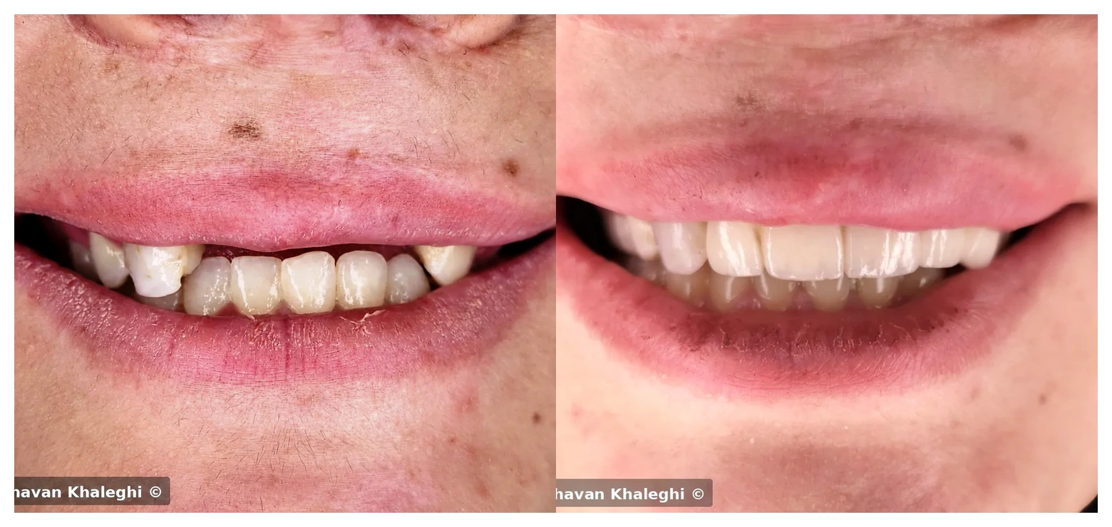
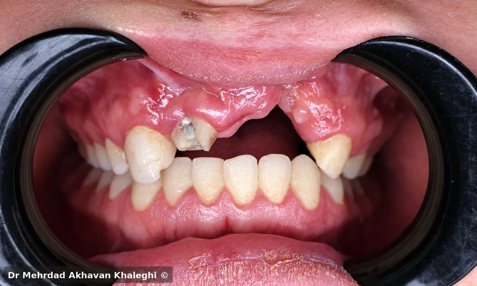
In a patient with a cleft, the position, form, and spacing of the teeth cannot always be expected to follow the pattern of a conventional dental arch. In this patient, tooth eruption, the available mesiodistal space, and the soft-tissue anatomy were atypical. If the design had been forced to follow tooth numbers or an ideal midline, the pontics would have required unnatural widths and proportions.
The central question was not how to replace three missing teeth one-for-one. It was how to distribute the space that actually existed so the replacement teeth would look natural together, remain in proportion with the existing dentition and facial tissues, and still provide function and maintainability.
Proportion versus symmetry
When a perfect midline would make the result less natural
In smile design, alignment of the dental and facial midlines—or at least a balanced relationship between them—is usually desirable. In this patient, however, the cleft-related anatomy and unusual distribution of space meant that perfect midline alignment could only be achieved at the cost of a different compromise.
If the midline had been moved toward an ideal position at any cost, the mesiodistal dimensions of the pontics would have had to be altered disproportionately. Some teeth would have become excessively wide and others excessively narrow, producing a visual discrepancy that could have been more obvious than the limited midline discrepancy itself.
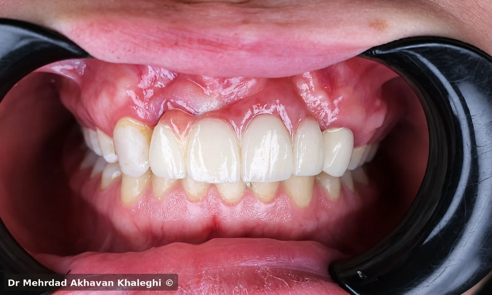
The decision was therefore to prioritize overall proportion rather than pursue geometric symmetry at any cost. The dimensions and positions of the replacement teeth were designed according to the available space, the adjacent teeth, and their relationship to the smile and facial tissues.
That became one of the important lessons of this case: something that looks “asymmetric” on paper may appear far more natural in the patient’s face and smile than symmetry forced onto anatomy that cannot support it.
Preparing the abutment teeth
A defensible foundation before a long-span reconstruction
Teeth 12 and 23 were not included in the plan simply because they happened to border the edentulous area. Both teeth independently required restorative treatment because of structural damage and previous treatment, while still providing adequate periodontal support and ferrule for use as abutments. Both also had inadequate previous root-canal treatment and were retreated before the prosthetic phase.
Tooth 12 required a post-and-core because of extensive structural loss, while tooth 23 was restored with a fiber post and composite build-up. Using these teeth as abutments therefore did not mean preparing two healthy teeth solely to create a bridge.
Bridge design
Esthetics without sacrificing maintainability
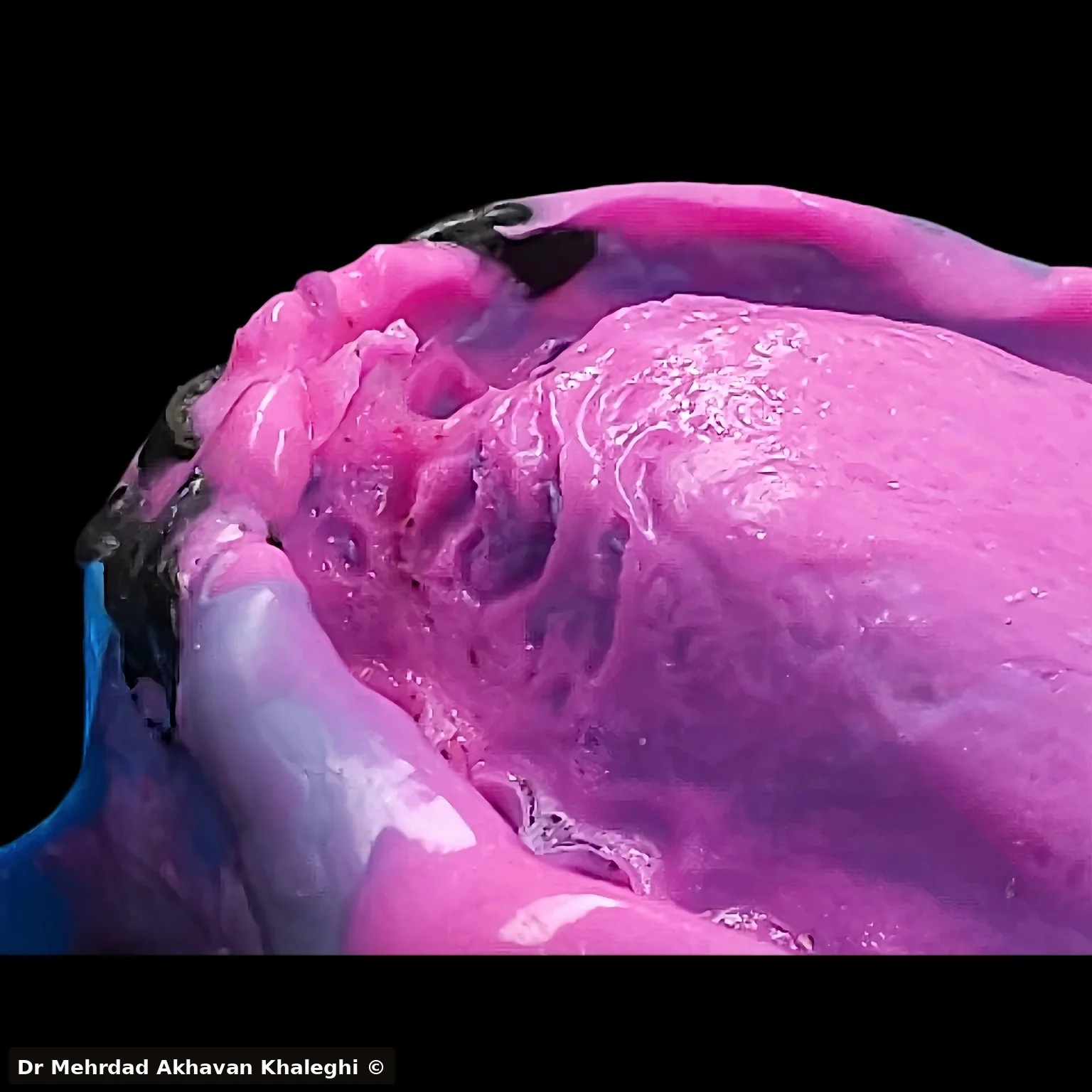
The definitive bridge was fabricated in zirconia, with cut-back and ceramic layering limited to the buccal surface. This allowed control of the esthetic surface while preserving the zirconia framework on the remaining aspects.
In a long-span FDP, however, appearance is only part of the problem. The three pontics occupied a region with atypical soft-tissue anatomy, and the patient also needed to be able to clean beneath the bridge over the long term.
The tissue surfaces of the pontics were designed to be convex, with enough mucosal contact to create a natural form without unnecessary tissue pressure. At the same time, access for flossing and cleaning beneath the pontics was maintained.
This part of the design was as important as the shape of the teeth. A bridge may look excellent on the day of delivery, but if the patient cannot keep it clean, the design was incomplete from the beginning.
Occlusion
Protecting the abutments in a long-span bridge
The occlusal design also had to acknowledge that forces applied to the pontics would ultimately be transferred to the two abutment teeth.
The pontics were kept out of contact in MIP to reduce direct and continuous loading of the pontics and, consequently, the abutments.
During left lateral movement, tooth 23—the patient's natural canine and one of the bridge abutments—maintained contact and contributed to posterior disclusion. In protrusive movement, contacts were kept light and controlled so that the movement could be guided without imposing unnecessary force on the bridge or its abutments.
The aim was not to remove the prosthesis completely from function; it was to ensure that every occlusal contact had a specific reason.
Treatment outcome
Harmony instead of forced symmetry
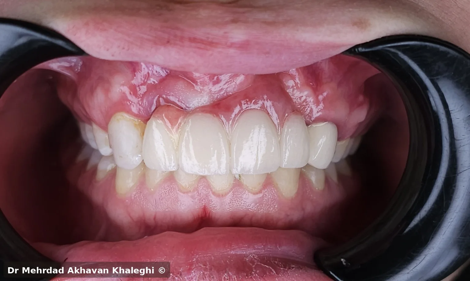
In the definitive result, perfect alignment of the dental and facial midlines was not possible, but the tooth proportions, their relationship to the existing dentition, and the overall harmony of the smile were preserved. Rather than expressing the spatial discrepancy through several unnaturally proportioned pontics, the available space was divided in a way that created a more balanced visual result.
At the same time, the pontic form preserved hygiene access, and the occlusal scheme was adjusted to reduce unnecessary loading of the abutments.
Four-year follow-up
An outcome that cannot be judged only on delivery day
The patient was followed clinically for approximately four years. No clinically relevant complication was reported during this period, and the tissue beneath the pontics remained free of signs of inflammation.
This follow-up mattered because the result of this case could not be judged only from a photograph taken on the day of delivery. The tissue design of the pontics, the patient's ability to clean the prosthesis, and the behavior of the abutment teeth over time were part of the real success of the treatment.
Conclusion
Symmetry alone is not the goal of treatment
The value of this case was not simply that a missing space could be restored with a five-unit bridge. The more important point was that the patient's atypical anatomy was not forced to obey the rules of a conventional dental arch.
In this patient, insisting on a perfectly ideal midline could have produced teeth with unnatural dimensions. The more defensible choice was to accept the existing limitation and, within that limitation, create the best possible balance among tooth form, smile, function, hygiene, and biologic conditions.
Symmetry alone is not the goal of treatment; the goal is to create harmony that is natural, functional, and maintainable within the actual conditions of that patient.
Clinical case and photography © Dr Mehrdad Akhavan Khaleghi. Unauthorized use or reproduction prohibited.