Clinical Case 3 · Supporting
Fixed Implant-Supported Rehabilitation of Both Arches: When the Value of Design Extends Beyond the Day of Delivery
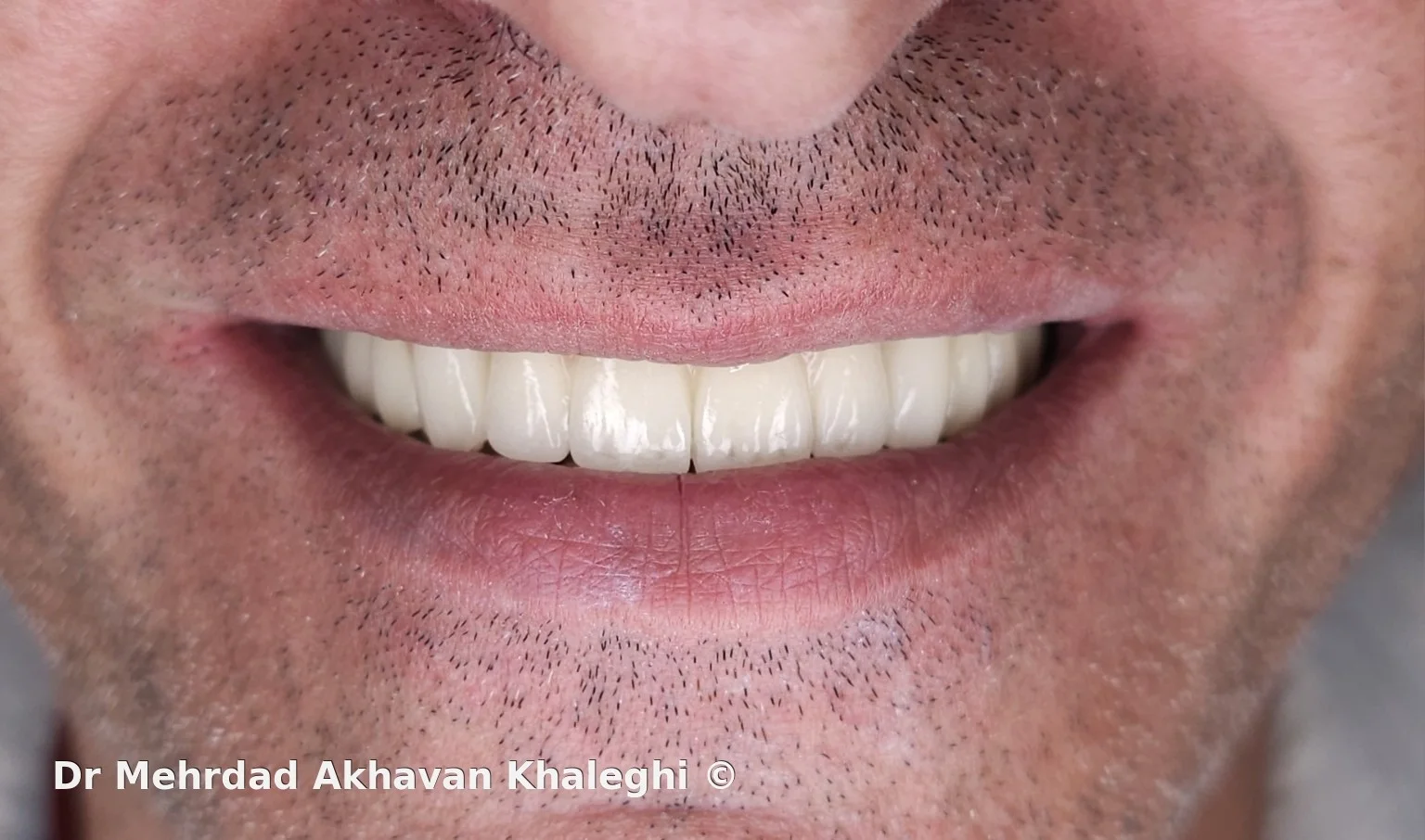
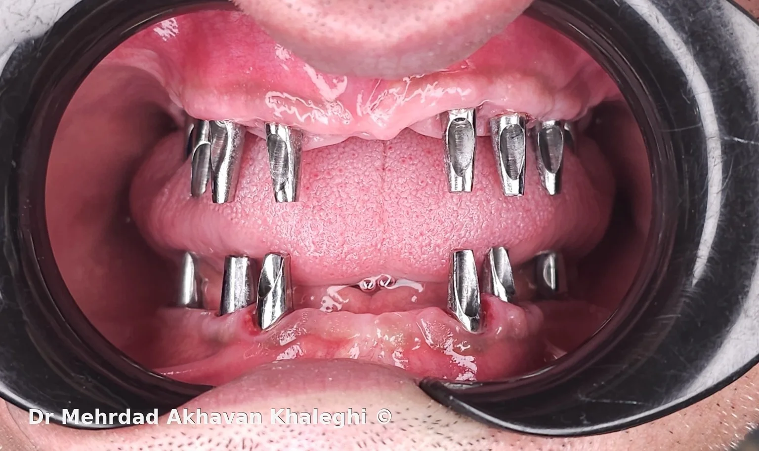
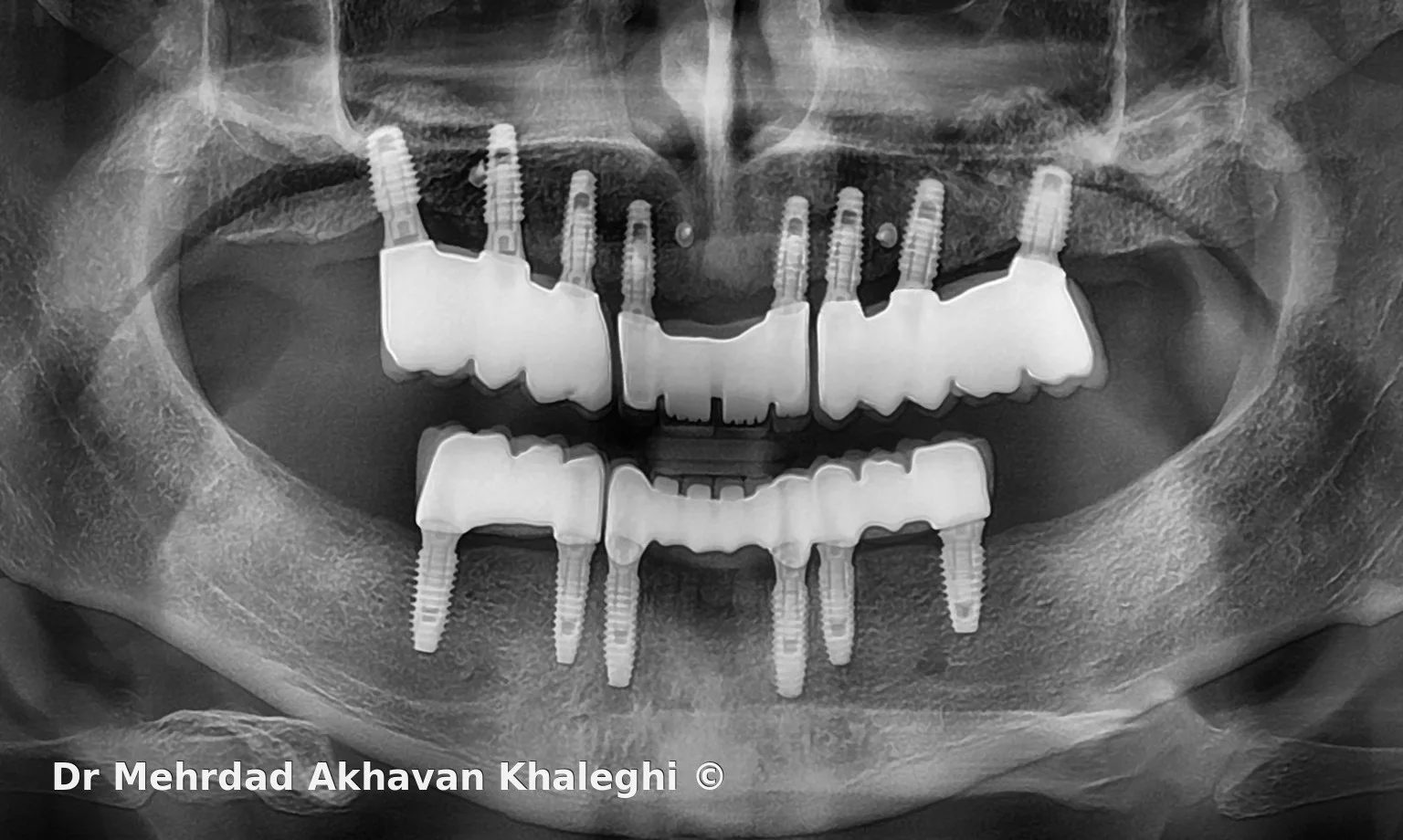
The treatment plan had been established before the patient reached complete edentulism. The patient had no history of wearing removable prostheses and, after the treatment options were reviewed, expressed a clear preference for a fixed implant-supported reconstruction. The edentulous ridges were favorable, and no major functional limitation prevented this approach. Eight maxillary implants and six mandibular implants ultimately formed the foundation of the rehabilitation.
In full-arch treatment, implant number is only one part of the problem. A more important question is how much of the proposed result can be evaluated clinically before definitive fabrication—and how much of the treatment can later be documented and judged over time.
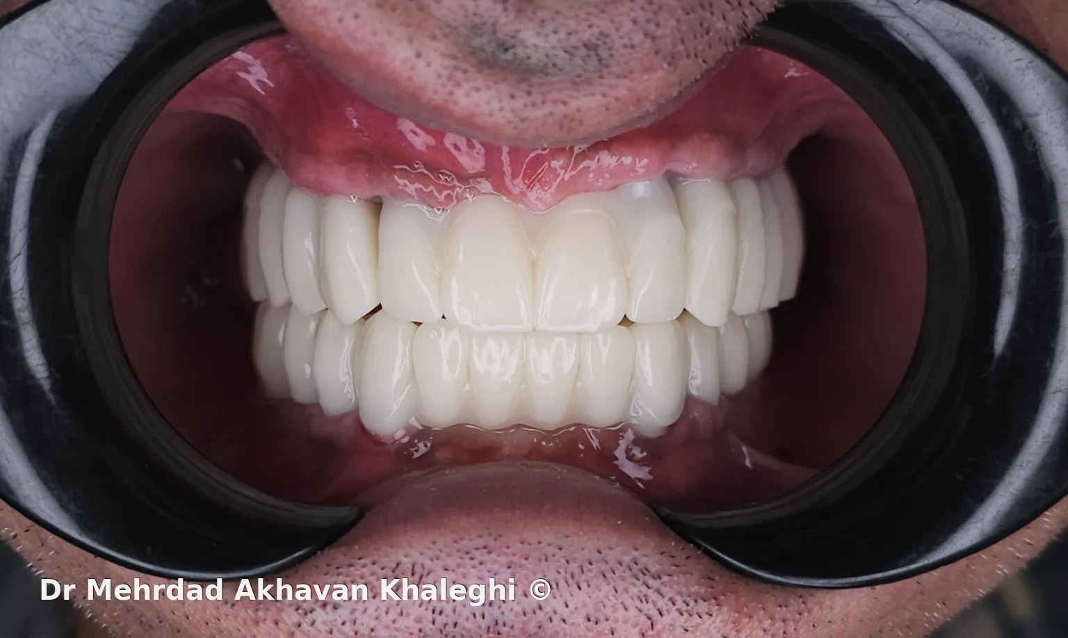
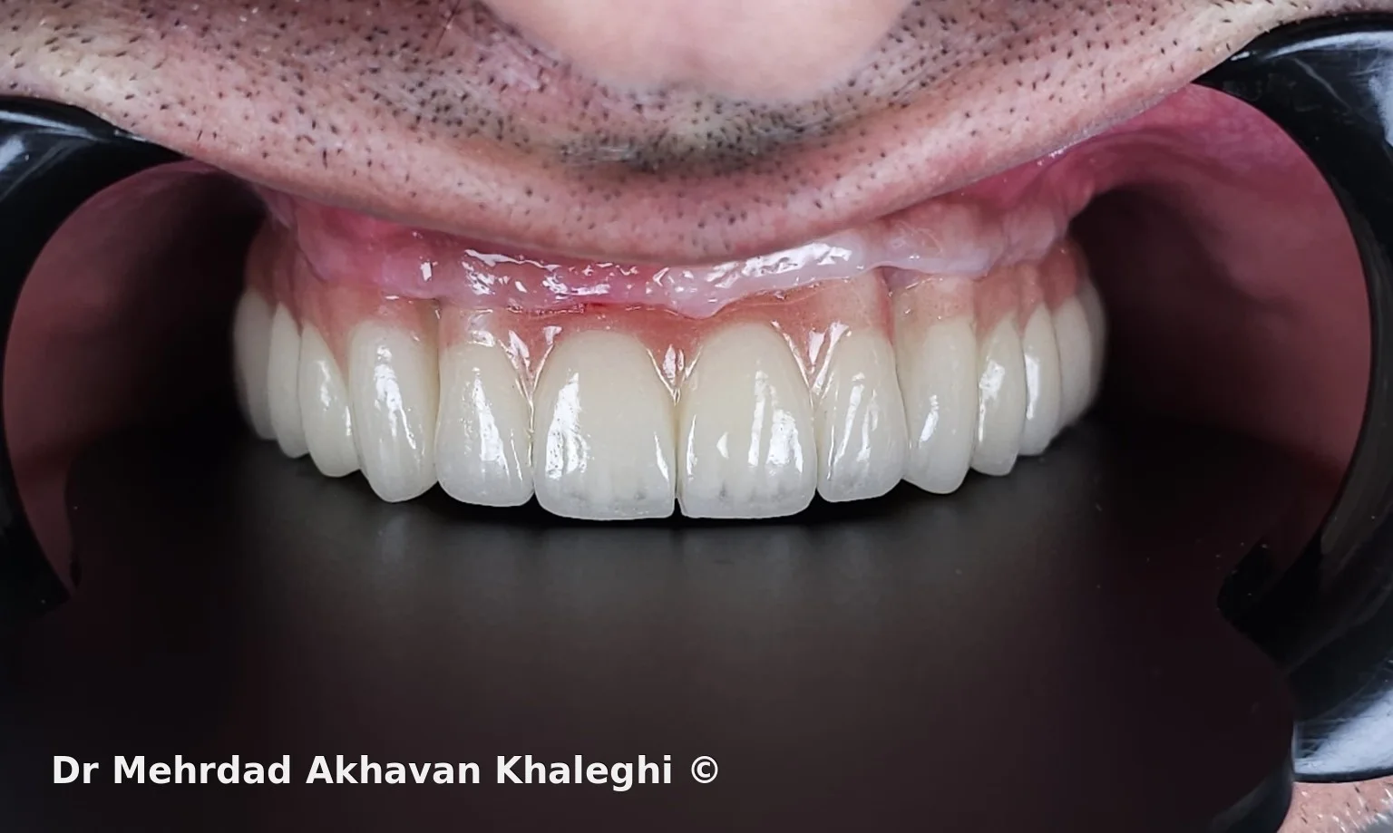
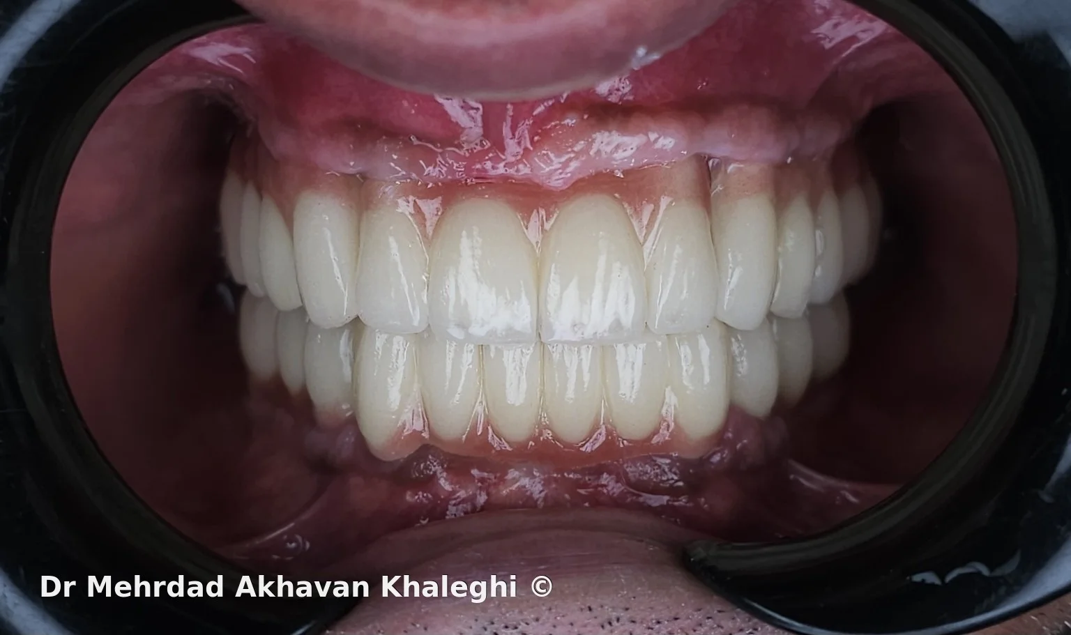
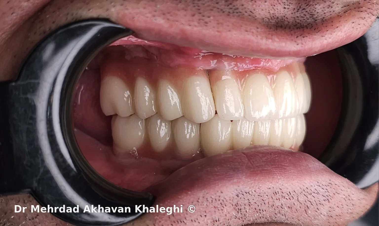
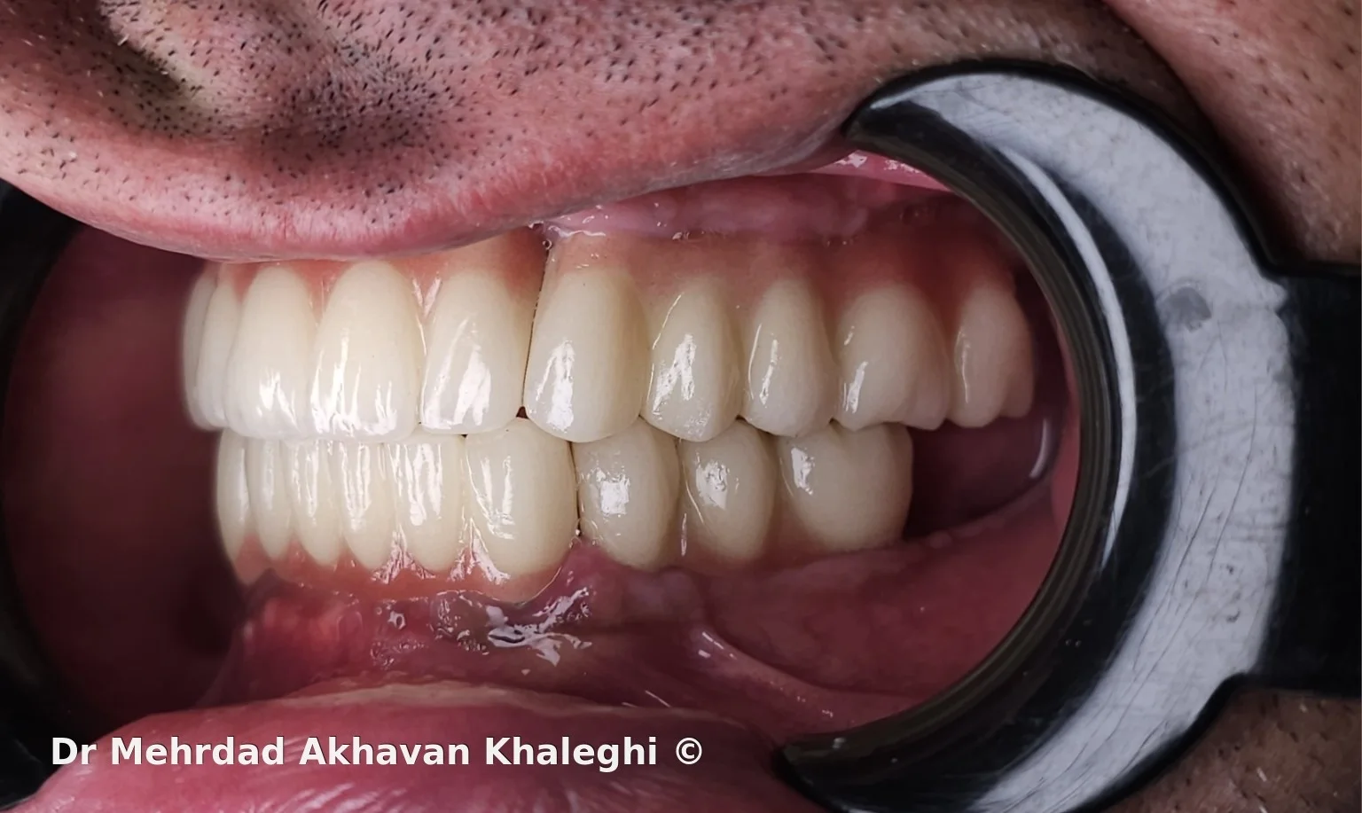
The definitive reconstruction was segmented rather than fabricated as one continuous prosthesis in each arch: four independent segments in the maxilla and three in the mandible. The restorations were cement-retained, with zirconia frameworks and ceramic layering.
Some of the most important decisions, however, were evaluated before the definitive restorations were made.
Before definitive fabrication: while the treatment could still be tested and changed
In a fixed rehabilitation of both arches, tooth position, vertical dimension, maxillomandibular relationships, occlusion, and perioral support all influence the final result. The more extensive the reconstruction, the more difficult and costly it becomes to correct a poor decision after definitive fabrication.
For that reason, acrylic provisional restorations were used to evaluate the proposed design clinically before proceeding to the definitive stage.
Vertical dimension and jaw relationships, tooth position, lip support, smile line, and the patient's return to function could all be assessed during this phase. A photograph of the provisionals remains part of the case documentation and records a point at which the result had not yet become definitive and could still be evaluated and modified.
The value of a provisional in this setting is not simply to provide a temporary restoration. In an extensive reconstruction, it can reveal the difference between what appears appropriate on a model or in a design and what actually works in the patient's mouth.
Once acceptable conditions had been established, information from the provisional phase became one of the references for definitive fabrication. The final tooth form and position and the functional relationships were therefore not created from zero; part of the proposed result had already been tested clinically.
Why does a full arch have to be one piece?
Maxilla
8 implants · 4 segments
Mandible
6 implants · 3 segments
After the provisional phase had established acceptable clinical conditions, another question remained: how should the definitive reconstruction be designed so that it would remain manageable in the future?
A one-piece restoration can appear conceptually simpler in an extensive reconstruction, but a localized problem may then involve a much larger portion of the treatment.
In this case, the maxillary reconstruction was divided into four segments and the mandibular reconstruction into three. Implant distribution and the relatively wide spacing between some fixtures also contributed to the decision to segment the reconstruction. If removal, repair, or remake is ever required, intervention does not necessarily have to involve the entire arch.
This is not a general claim that segmentation is superior to a one-piece design. Implant position and distribution, span length, prosthetic space, path of insertion, restorative materials, and the conditions of the individual patient all need to be considered when selecting the design.
The broader principle is that maintainability is better considered at the time of planning than only after a complication occurs.
An extensive reconstruction without making it inseparable
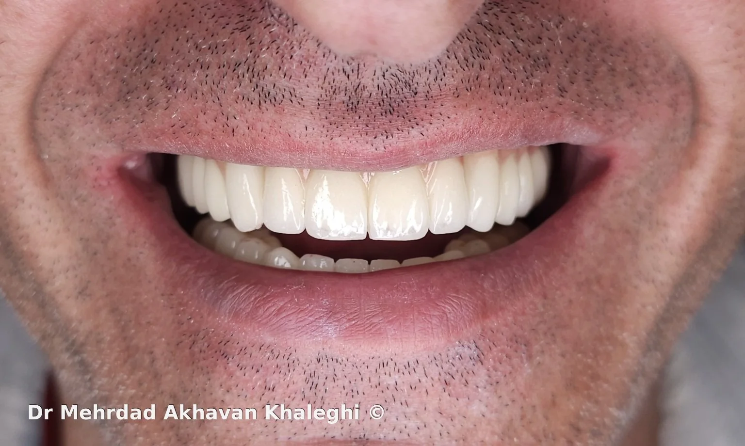
Although the definitive prostheses were divided into multiple segments, they still had to function visually and occlusally as one coordinated reconstruction. Segmentation should not come at the expense of continuity in the final result.
The restorations were cement-retained. Cement retention, like any retention strategy, has advantages and limitations and requires careful control of cementation and removal of excess cement.
Its use here should not be read as evidence that cement retention is superior to screw retention. In implant-supported treatment, retention mode must be considered alongside implant position, restorative design, retrievability, and future maintenance.
Layered zirconia and the limits of judging a case at delivery
The definitive restorations were fabricated with zirconia frameworks and ceramic layering.
In a full-arch reconstruction, an acceptable esthetic result on the day of delivery is only part of the assessment. Restoration volume, ceramic support, occlusion, force distribution, and material behavior under function cannot be judged completely from the delivery appointment alone.
Some prosthetic or biologic problems, if they occur, may only become apparent months or years later. Follow-up documentation is therefore part of the treatment record rather than simply an opportunity to obtain another final photograph.
The two-year radiograph: one documented point in the follow-up pathway
A radiograph from approximately two years after treatment is also available for this patient.
Its value is straightforward: the rehabilitation is not documented only by images taken at delivery. At least one later point in the course also has radiographic documentation.
A single radiograph at two years cannot describe the entire biologic behavior of the implants over a five-year period, and it should not be asked to prove more than it can show. Its importance here is as one part of a documented follow-up pathway—a later point that can be compared with the treatment record rather than relying only on the initial result.
Approximately five years later
Follow-up continued for approximately five years, during which no significant prosthetic or implant complication was reported.
That is a favorable outcome, but a single case cannot establish that implant number, the provisional phase, segmentation, retention mode, or layered zirconia was independently responsible for it. Long-term behavior of an extensive reconstruction reflects a combination of surgical, prosthetic, occlusal, biologic, and maintenance-related factors.
Likewise, the absence of a complication does not make planning for maintenance irrelevant. At the time of fabrication, it is impossible to know which patient may later require a repair or modification. The ability to intervene in the future is part of planning even when that ability ultimately never has to be used.
For me, the value of this case lies across three points in time: first, the proposed result could be observed and evaluated in the provisionals; then the definitive reconstruction was delivered; and later, part of its behavior was documented through clinical follow-up and an approximately two-year radiograph, with follow-up extending to about five years.
None of those stages alone proves treatment success. Together, however, they provide a more complete picture of the rehabilitation than a simple before-and-after comparison.
In an extensive reconstruction, judgment should not be confined to the day of delivery. What can be tested before definitive fabrication should be tested, and what can only be understood with time should be followed. The value of a design is complete only when the treatment is not merely deliverable, but also monitorable and manageable over the long term.
Clinical case and photography © Dr Mehrdad Akhavan Khaleghi. Unauthorized use or reproduction prohibited.